This article is informational only and not medical advice. It summarizes how major guidelines and reviews discuss imaging (MRI/CT) in facial nerve palsy.
TL;DR:
In a typical new-onset Bell’s palsy, routine MRI/CT is not recommended, especially within the first ~3 months after diagnosis. Imaging is considered when the presentation is atypical or recovery is absent.
MRI is preferred for suspected nerve/brain/parotid pathology; high-resolution temporal bone CT is preferred for trauma and bony middle-ear disease (e.g., cholesteatoma).
When imaging is not routinely recommended
For a first episode of idiopathic peripheral facial nerve palsy with a classic, sudden onset and no concerning features, leading guidelines advise against routine MRI/CT. Instead, clinicians monitor early recovery and reassess if the course is atypical.
Red flags: when to consider MRI/CT
Consider imaging (and specialist referral) if any of the following are present:
No clinical improvement over time - Particularly if no meaningful recovery by ~3 months, reassessment (often including imaging) is advised.
Recurrent facial palsy or unusual distribution (limited to one branch) - Raises suspicion for non-idiopathic causes.
Other cranial nerve/neurologic symptoms - e.g., new hearing loss, , , diplopia, dysphagia, limb weakness or sensory loss. These are for Bell’s palsy and warrant further work-up.
Share article:
Join the Waitlist!
You'll be the first to know and one of our earliest testers.
🎁
Join to get a free e-book
Practical guide for patients with facial nerve paralysis
100K+
NEED SUPPORT
annually worldwide
tinnitus
true vertigo
not typical
Ear disease or temporal bone concerns - Chronic ear disease or suspected cholesteatoma; temporal bone trauma. In these settings, high-resolution CT of the temporal bone is emphasized.
Ramsay Hunt syndrome (herpes zoster oticus) or severe otologic symptoms - Presentation and management differ from Bell’s palsy; atypical courses may prompt imaging.
Suspicion of neoplasm - Progressive weakness, palpable parotid mass, or angle-of-the-cerebellopontine-junction symptoms. MRI with contrast helps evaluate the facial nerve, CP angle, and parotid.
Bilateral facial palsy or concern for central (brain) causes - Requires a broader differential and often imaging/urgent assessment.
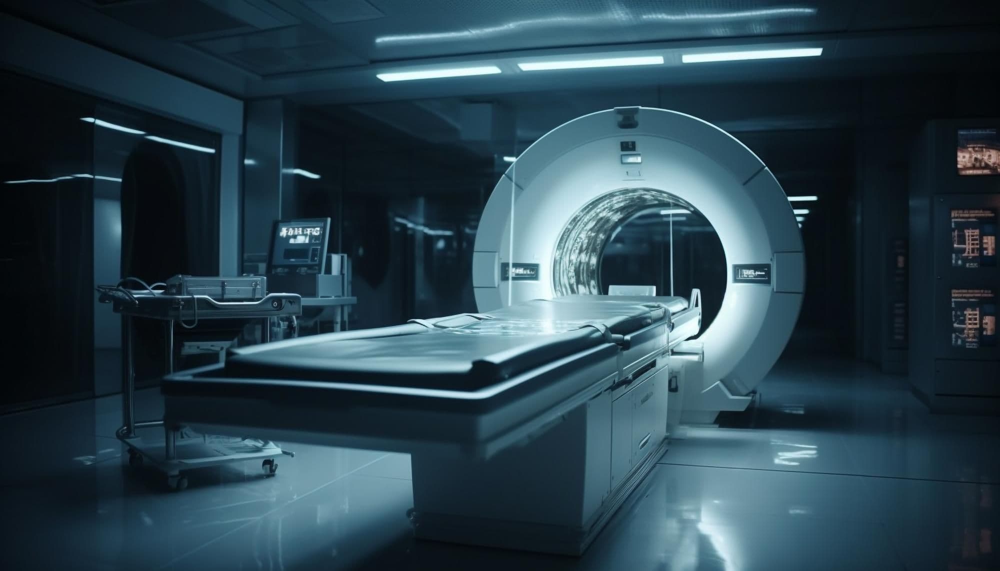
MRI vs CT: which test and why?
MRI (with contrast)
Best for the facial nerve along its course, brainstem/CPA, and parotid. Helpful when the exam suggests neuritis, intracranial pathology, or tumor; also used when recovery stalls.
High-resolution CT (temporal bone)
Best for bony anatomy: fractures, ossicular chain, dehiscence, and chronic ear disease/possible cholesteatoma. Often first-line when trauma is suspected.
What can imaging show in Bell’s palsy?
In Bell’s palsy, contrast enhancement of involved facial-nerve segments on MRI can be seen, reflecting neuritis/inflammation. These findings support-but do not define-the diagnosis, which remains clinical in typical cases.
“Should everyone get an MRI just in case?”
A 2023 French multicenter cohort reported that routine MRI reclassified a small minority of presumed Bell’s palsy cases by revealing alternative causes—provoking discussion about broader imaging—but this is not current consensus and contrasts with guideline advice to avoid routine imaging.
FAQ
Does imaging confirm Bell’s palsy?
Not by itself. Bell’s palsy remains a clinical diagnosis; MRI may show nerve enhancement but is not routinely required when the presentation is typical.
If I have persistent or worsening symptoms, is imaging considered?
Yes-lack of improvement by ~3 months, recurrence, additional neurologic signs, trauma, ear disease, or tumor concern are common indications to image.
Which modality has radiation?
CT uses ionizing radiation; MRI does not. Choice depends on the suspected cause (nerve/soft tissue vs. bone/trauma).
How NeuroFace can help
The NeuroFace app was created to support people with facial nerve palsy in rehabilitation. Thanks to a simple mobile interface:
It offers facial expression exercises that can be done at home.
It enables progress monitoring - the app records exercise sessions and helps observe changes in facial expression over time.
It reminds users to perform regular exercises and educates them about eye hygiene, skin care and safe rehabilitation practices.
It collaborates with specialists - data from the app can be shared with doctors and physiotherapists, allowing better tailoring of therapy.
NeuroFace does not replace a medical visit, but it can be a valuable complement to professional rehabilitation. Our goal is to make daily work on facial expression easier for patients and provide motivation for regular exercises.
Sources
AAO-HNSF – guidelines and summary (no routine imaging diagnostics; reassessment if no improvement after 3 months). entnet.org
AAO13 (2024) – quality measure: do not routinely imager in 3 months, exceptions = atypical features (list of ‘denominator exceptions’). entnet.org
NICE CKS – in typical form without routine imaging; indications for urgent referrals in case of red flags. cks.nice.org.uk , cks.nice.org.uk
RACGP – red flags atypical for Bell's palsy (e.g. diplopia, dysphagia, true numbness, dizziness). racgp.org.au
ACR Appropriateness and radiological examinations – MRI for the nerve/CNS/parotid gland; HRCT for the temporal bone/middle ear/injury. jacr.org, pmc.ncbi.nlm.nih.gov
JAMA Network Open 2023 – cohort suggesting the benefit of routine MRI (position different from AAO-HNSF). JAMA Network
 Author: Designed by Freepik